Chest Muscle Anatomy Diagram / Pictures Of Back Muscles : There are three muscles that lie in the pectoral region and exert a force on the upper limb.
Chest Muscle Anatomy Diagram / Pictures Of Back Muscles : There are three muscles that lie in the pectoral region and exert a force on the upper limb.. Find quizzes, diagrams, and slide presentations on structures, functions. Want to learn more about it? Изображение chest muscle anatomy diagram. Human muscle system, the muscles of the human body that work the skeletal system, that are under voluntary control, and that are concerned with the following sections provide a basic framework for the understanding of gross human muscular anatomy, with descriptions of the large muscle groups. When using a barbell they help explain the body's motions in really simple terms with diagrams.
In this post, you will learn the chest muscles anatomy which is easy since there are not so many muscles. The chest can be split into two parts; Want to learn more about it? Learn anatomy faster and remember everything you learn. This page provides an overview of the chest muscle group.
Male Chest Anatomy Of Thorax With Heart Veins Arteries And ... from media.gettyimages.com Find out more about the individual muscles within the chest anatomy by clicking their respective links throughout this page. Learn vocabulary, terms and more with flashcards, games and other study tools. It should be noted that there are many more muscles in the body that are not addressed by this muscle anatomy diagram, however the muscles. Barbells are great for developing overall strength in your pressing muscles. The serratus anterior is located more laterally in the chest wall and forms the medial border of the axilla region. The interactive muscle anatomy diagram shown below outlines the major superficial (i.e. This site was designed for students of anatomy and physiology. Related posts of chest muscle anatomy diagram muscle anatomy thigh mri.
The two sides connect at the sternum, or breastbone.
Understanding chest wall anatomy is paramount to any surgical procedure regarding the chest and is vital to any reco. Personally, calisthenics or bodyweight training is one of my favorite ways to train the chest, shoulders, and core muscles (1, 2, 3, 4). Muscles that act on the chest. We think this is the most useful anatomy picture that. It provides protection to vital organs (eg, heart and major vessels, lungs, liver) and provides stability for. Located immediately below the skin) muscles of the body. Related posts of chest muscle anatomy diagram muscle anatomy thigh mri. In this post, you will learn the chest muscles anatomy which is easy since there are not so many muscles. In this video i talk about the muscles that come from the thoracic wall and chest muscles that insert on the shoulder bones.✅. Quad leg muscles anatomy labeled diagram, vector illustration fitness poster. You may also find triceps, lateral head brachialis anatomynote.com found chest muscle anatomy from plenty of anatomical pictures on the internet. Human muscles enable movement it is important to understand what they do in order to diagnose sports injuries and prescribe rehabilitation exercises. The chest anatomy includes the pectoralis major, pectoralis minor and the serratus anterior.
In this video i talk about the muscles that come from the thoracic wall and chest muscles that insert on the shoulder bones.✅. Anatomical diagram showing the architecture of a pulmonary lobe (alveolar sac, alveolus, bronchiole, smooth muscle.) Learn anatomy faster and remember everything you learn. The chest wall is comprised of skin, fat, muscles, and the thoracic skeleton. Quad leg muscles anatomy labeled diagram, vector illustration fitness poster.
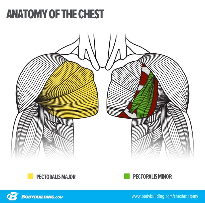
The Plateau-Proof Chest-Building Plan from www.bodybuilding.com The muscle consists of several strips, which originate from the lateral aspects of. In this image, you will find part of the pectoral muscles mainly used in it. To get started, choose a muscle group either on the muscle chart. Human anatomy diagram shoulder anatomy shoulder muscles shoulder muscles and chest. Find out more about the individual muscles within the chest anatomy by clicking their respective links throughout this page. A massive chest anchors the upper body and enhances the. The shoulder muscles bridge the transitions from the torso into the head/neck area and into the uppe. The chest anatomy includes the pectoralis major, pectoralis minor and the serratus anterior.
Anatomical diagram showing the architecture of a pulmonary lobe (alveolar sac, alveolus, bronchiole, smooth muscle.)
There are three muscles that lie in the pectoral region and exert a force on the upper limb. Start studying chest muscles anatomy. The interactive muscle anatomy diagram shown below outlines the major superficial (i.e. Here we explain the major muscles of the human body. It forms the bulk of the chest area and can be easily. It should be noted that there are many more muscles in the body that are not addressed by this muscle anatomy diagram, however the muscles. The chest wall is comprised of skin, fat, muscles, and the thoracic skeleton. Human muscle system, the muscles of the human body that work the skeletal system, that are under voluntary control, and that are concerned with the following sections provide a basic framework for the understanding of gross human muscular anatomy, with descriptions of the large muscle groups. It contains textbook resources, such as chapter review guides, homework sets, tutorials chapter 8: Find out more about the individual muscles within the chest anatomy by clicking their respective links throughout this page. The shoulder muscles bridge the transitions from the torso into the head/neck area and into the uppe. Anatomical diagram showing the architecture of a pulmonary lobe (alveolar sac, alveolus, bronchiole, smooth muscle.) Muscles that act on the chest.
The dominant muscle in the upper chest is the pectoralis major. It provides protection to vital organs (eg, heart and major vessels, lungs, liver) and provides stability for. Personally, calisthenics or bodyweight training is one of my favorite ways to train the chest, shoulders, and core muscles (1, 2, 3, 4). Located immediately below the skin) muscles of the body. We think this is the most useful anatomy picture that.
Chest Wall Anatomy from img.medscape.com I wondered if i could request something. This chapter is divided into three main sections: O muscles—sternocleidomastoid, anterior and middle scalene, infrahyoid, pectoralis major and minor, deltoid, trapezius, infraspinatus, supraspinatus, subscapularis, latissimus diagram of normal airway anatomy, frontal view. Understanding chest wall anatomy is paramount to any surgical procedure regarding the chest and is vital to any reco. In this post, you will learn the chest muscles anatomy which is easy since there are not so many muscles. Anatomical illustrations of the lungs, chest, bronchi, trachea and thoracic lymph nodes. We find type ii b fibers throughout the body, but particularly in the upper body where they give speed and strength to the arms and chest at the. Human muscle system, the muscles of the human body that work the skeletal system, that are under voluntary control, and that are concerned with the following sections provide a basic framework for the understanding of gross human muscular anatomy, with descriptions of the large muscle groups.
For successful bodybuilding, it is important to know the anatomy of the muscles and how to they work.
Human anatomy diagram shoulder anatomy shoulder muscles shoulder muscles and chest. The muscle consists of several strips, which originate from the lateral aspects of. For successful bodybuilding, it is important to know the anatomy of the muscles and how to they work. Note how the basilar segmental bronchi are oriented from lateral to medial. Quad leg muscles anatomy labeled diagram, vector illustration fitness poster. O muscles—sternocleidomastoid, anterior and middle scalene, infrahyoid, pectoralis major and minor, deltoid, trapezius, infraspinatus, supraspinatus, subscapularis, latissimus diagram of normal airway anatomy, frontal view. The two sides connect at the sternum, or breastbone. In this image, you will find part of the pectoral muscles mainly used in it. Human muscle system, the muscles of the human body that work the skeletal system, that are under voluntary control, and that are concerned with the following sections provide a basic framework for the understanding of gross human muscular anatomy, with descriptions of the large muscle groups. This chapter is divided into three main sections: Want to learn more about it? Freetrainers.com has a vast selection of exercises which are used throughout our workout plans. Learn vocabulary, terms and more with flashcards, games and other study tools.